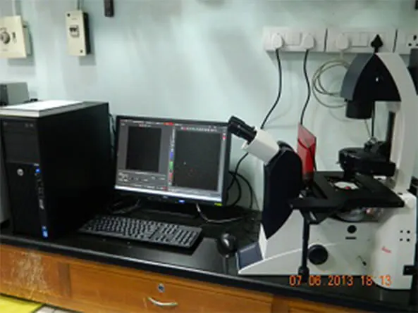
Hyperspectral Imaging
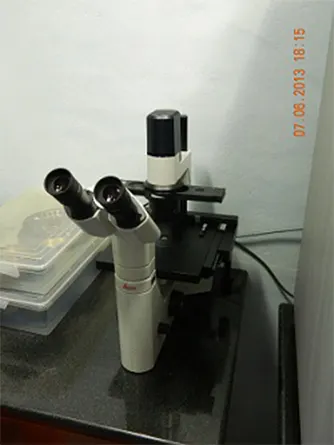
Instrument Name: The CytoViva� Hyperspectral Imaging System
Available magnifications: 10X and 40X air objectives, 60X and 100X oil immersion objectives
Spectral range: 400-1000 nm
Light sources:
Halogen light source (400-750 nm)
Exfo X-Cite 120 fluorescent light source with excitations for DAPI, FITC and TEXAS RED dyes
Instrument Description:
Hyperspectral Imaging System (HSI) provides spectral analysis of materials and biologicals imaged with the nano-scale optical microscope operating in the visible near infrared spectral range (VNIR). It supports research in areas such as nano-drug delivery and nano-toxicology. This spectral analysis method supports both non-fluorescent and fluorescently labeled components in live cells and nano-materials.
This nanoscale microscope combined with dark field condenser, motorized stage and HSI system provides analytical confirmation of nano-scale materials and their interactions with biologicals or other composite materials.The spectrophotometer attached with this system contains original, aberration-corrected convex holographic diffraction gratings which provides superior signal-to-noise ratio. HSI analysis software (ENVI) enables the researcher to easily locate regions within the sample that match qualified spectra that has been preloaded in a library. It also provides the ability to un-mix spectra to isolate specific sample components or sample anomalies for further evaluation.