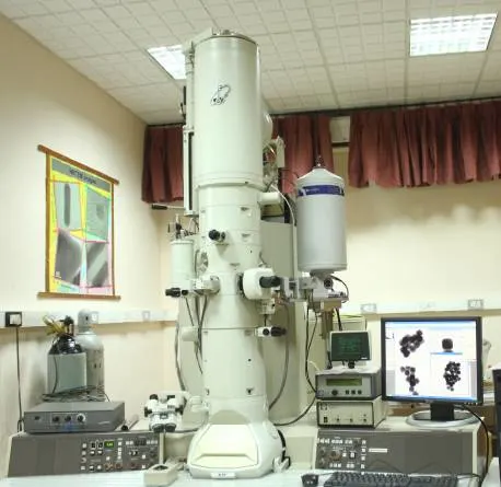
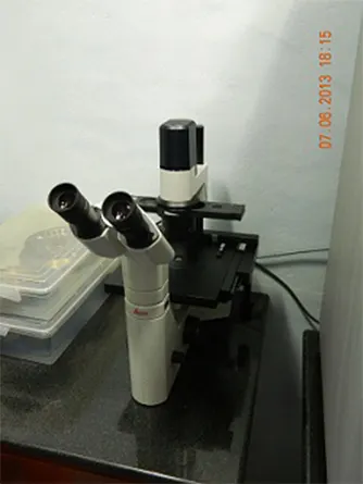
Inverted Fluorescence Microscope
Available magnifications: 10X, 20X, 40X and 63X air objectives, 100X oil immersion POL objective.
Camera: Leica DFC365 FX.
Light source: Halogen light source (12 V, 100 W)
Filter cubes: Leica filter cubes, A (UV), I3 (blue) and N2.1(green).
Software: Leica AF6000
Instrument description:
Leica DMI3000 B is an inverted microscope and can be used for fluorescence imaging of materials and biological samples. This inverted fluorescence microscope is combined with phase contrast microscopy and dark field imaging at 10X, 20X magnification which can be further used for analytical confirmation of nano-scale materials and their interactions with biologicals or other composite materials. Three different filter cubes A (UV), I3 (blue) and N2.1 (green) provide excitation from UV-green region and can be used for excitation of multiple dyes. Leica AF6000 software with time lapse module can be used for imaging of samples in dynamic environment. It supports research in multiple areas such as fluorescent material’s characterization bio and-labelling. Imaging analysis method can be used for fluorescently labelled components in live cells and nano-materials.