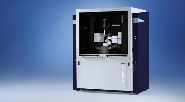
Single Crystal X-ray Diffractometer
Instrument specification
Bruker Single Crystal X-ray Diffractometer (SCXRD), D8 Venture with Photon III C14 detector, is used to determine three dimensional crystal structures. It is equipped with two micro-focus X-ray sources (Cu and Mo radiation) providing a choice of two wavelengths, for measuring patterns in very small crystals. Powder diffraction of capillary samples can also be obtained. Grazing incidence measurements can also be made. Different cooling systems are available, enabling low temperatures such as 100 K.

Technicalities
Key features of D8 Venture SCXRD
Flux density: 6 × 10¹¹ X-rays/mm2s
X-ray sources: IµS 3.0 and IµS DIAMOND microfocus, METALJET liquid metal
Active area: up to 280 cm2
Anode: Microfocus TXS Rotating Anode
Goniometer: FIXED-CHI or KAPPA, for completeness and multiplicity
Software and data processing: APEX5 and PROTEUM3
Low temperature range: Cryostream (liquid nitrogen) and COBRA (non-liquid nitrogen)
Theory of operation
A crystal has a pattern of unit cells repeated in a three-dimensional lattice. Upon X-rays incident on the crystal lattice, it behaves like a diffraction grating, producing a diffraction pattern. A beam of parallel monochromatic X-rays of a specific wavelength is used to obtain this diffraction. The analysis of this diffraction pattern leads to the structure and shape of the lattice units. With the phase angles and a good fit between experimental and calculated diffraction patterns.

