Confocal Raman Microscope

Instrument specification
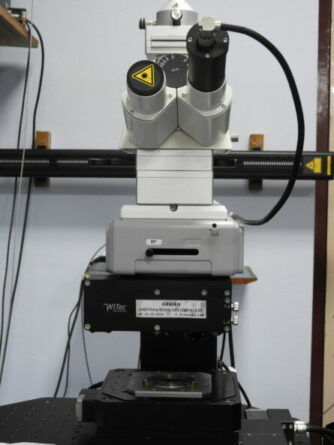
The confocal Raman Microscope, CRM alpha 300 S (WITec GmbH) is the best of its class instrument that combines in a unique way the operations of confocal microscopy, Raman spectroscopy, AFM and SNOM. This work station has been used for: (i) Confocal microRaman spectroscopy and microscopy (Raman spectrum for different varieties of samples such as solids, powders, thin films, liquid and so forth, SERS [air and liquid measurements], Raman spectral imaging), (ii) confocal section analysis, depth scanning in transmission and reflection modes, (iii) atomic force microscopy (contact and tapping modes), and (iv) aperture scanning near field optical microscopy (SNOM).
The confocal setup reduces unwanted background signals, enhances contrast and provides depth information. Differences in chemical composition, although completely invisible in the optical image, will be apparent in the Raman image and can be analyzed with a resolution down to 200 nm. Its sensitive setup allows for the nondestructive imaging of chemical properties without specialized sample preparation. With AFM, investigation of material properties on the nanometer scale is possible. As SNOM requires only minimal sample preparation if any, it is ideally suited to quickly and effortlessly image the optical properties of a sample with resolution below the diffraction limit. Typical applications are found in nanotechnology, materials research, life sciences and others.
Technicalities
Manufacturer: WITec GmbH, Germany
Operating Modes:
Confocal Micro Raman Spectroscopy and Microscopy,
Atomic Force Microscopy (AFM), Scanning Near-field Optical Microscopy (SNOM)
Excitation LASERs:
1. Frequency doubled Nd:YAG dye LASER [maximum power output is 50 mW power at 532 nm]
2. Helium Neon [HeNe] LASER [35 mW output power at 633 nm]
Scan Stage: The sample scan stage is a linear piezo-driven feedback controlled one with a scan area of 50 x 50 μm2. There is also stepper motor driven large area scan stage of maximum scanning area 12.5 x 12.5 mm2.
Detectors: Single counting photomultiplier tube (PMT) and Peltier cooled charge coupled device (CCD) were used for high sensitive detection of photons.
Key features of HRTEM technology
- Spatial resolution beyond the diffraction limit (ca. 60 nm laterally)
- Unique patented SNOM sensors
- Ease of use in air and liquids
- Nondestructive, label-free imaging technique provides super-resolution microscopy with minimal, if any, sample preparation
- Upgradeable with confocal Raman imaging for correlative microscopy and near-field Raman imaging
- Three techniques always integrated within one instrument: confocal microscopy, AFM and SNOM
Schematic of the JEOL 3010 instrument*

For more information on the instrument visit www.witec.de
Theory of operation
About the Instrument:
Selected Data:


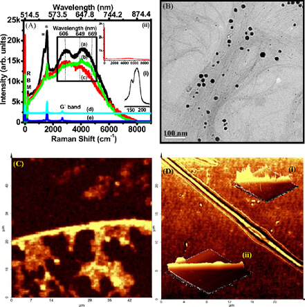
A) Single spot Raman spectra of (a) Ag-SWNT, (b) Au-SWNT, (c) AuNR-SWNT, (d) pristine SWNTs, and (e) SWNTs treated with heated citrate solution. Traces (d) and (e) are shifted vertically for clarity. Radial breathing mode (RBM) is labeled and the D and G bands are indicated by asterisks (*). The visible emission maxima are marked with their respective wavelengths. Inset (i): the RBM region at a higher resolution. Inset (ii): the Raman spectra of a dropcast film of AuNR, along with the Rayleigh to compare the intensities. (B) TEM image of the Au-SWNT. (C) Raman spectral images of AuNRSWNT, based on the intensities of visible emission in the 595 to 675 nm window. The spots observed away from the nanotube structure are due to shorter nanotubes remaining in the centrifugate or due to the z-axis discrimination of confocal imaging. (D) Light intensity based transmission SNOM image of AuNRSWNT. Inset (i): the three-dimensional view of (B), rotated suitably to show the increased emission. Inset (ii): corresponding topographic image. Raman and SNOM data are acquired with 514.5 nm excitation2

(a) Optical image of the AFM cantilever on a Random Access Memory (RAM)chip sample commonly used in personal computers. The squares marked in different colors, say red, blue and green corresponds to the AFM images labeled (b), (c)& (d) respectively. (e) Topography along the black line in the AFM image(b). (f) Topography along the red line in (d). (g) Non-contact mode AFM image obtained from a standard alumina pattern named Fischer pattern.
Footnotes:
1. http://www.witec.de/en/home
2. Using ambient ion beams to write nanostructured patterns for surface enhanced Raman spectroscopy, Anyin Li, Zane Baird, Soumabha Bag, Depanjan Sarkar, Anupama Prabhath, Thalappil Pradeep and R. Graham Cooks, Angew. Chem. Int. Ed., 2014, 53, 12528-12531
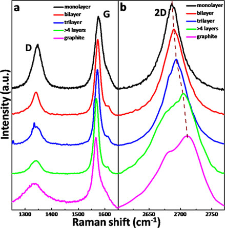
3. Single- and few-layer graphene growth on stainless steel substrates by direct thermal chemical vapor deposition, Robin John, Ashok Reddy, C. Vijayan and T. Pradeep, Nanotechnology, 2010, 22, 12528-12531.
4. Ambient electrospray deposition Raman spectroscopy (AESD RS) using soft landed preformed silver nanoparticles for rapid and sensitive analysis, Tripti Ahuja, Atanu Ghosh, Sandip Mondal, Pallab Basuri, Shantha Kumar Jenifer, Pillalamarri Srikrishnarka, Jyoti Sarita Mohanty, Sandeep Bose and Thalappil Pradeep, Analyst, 2019, 144, 7412-7420.
5. Organic solvent-free fabrication of durable and multifunctional superhydrophobic paper from waterborne fluorinated cellulose nanofiber building blocks, Avijit Baidya, Mohd Azhardin Ganayee, Swathy Jakka Ravindran, Kam Chiu Tam, Sarit Das, Robin Ras and Thalappil Pradeep, ACS Nano, 2017, 11, 11091−11099
 Search
 Sign in


 Total views : 241286